
What is Fibular Hemimelia?
Fibular hemimelia is a congenital anomaly involving the lower limbs. This condition is characterized by the absence of the fibula bone – this can range from partial (mild fibular hypoplasia) to complete (fibular aplasia). In general, this congenital appears on its own. However, in some cases, it can appear in association with the proximal focal femoral deficiency – this is defined by the partial absence of the proximal end of the femur (congenital anomaly as well). Fibular hemimelia is also found in association with the absence of phalanges in the lateral toes.
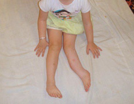
Picture of a Child with Fibular Hemimelia
The affectation is unilateral in the majority of the patients. It is also quite common for the postaxial portion of the limb to be affected, a condition which is known as paraxial fibular hemimelia. When the lateral rays of the foot are also affected, fibular hemimelia is presented as a complete terminal deficiency. There are patients in which the foot does not present any changes.
Epidemiology
Fibular hemimelia is considered a rare congenital anomaly. However, from all the congenital anomalies that involve the absence of certain bones, this is by far the most common. The incidence of fibular hemimelia is of 5.7 to 20 cases for every 1 million births. It is known that boys are more often affected by this condition than girls.
Signs and Symptoms of Fibular Hemimelia
The partial or complete absence of the fibula is noticed immediately after birth. The respective limb might appear shortened or a discrepancy can be noticed between the two limbs. Other changes that can be identified include: fusion of the toes (syndactyly), fewer toes than normal (oligodactyly) or more toes than normal (polydactyly).
The patient might present a fibrous band instead of the fibula, with the respective leg appearing short and deformed. The lateral part of the ankle joint can be absent, as the distal end of the fibula is missing. Because of that, the left part is unstable and the foot can also present deformities (equinovalgus). A part of the foot can also be absent – in such situations, the doctor can either decide to perform corrective surgery for bringing the foot into the normal function or perform amputation.
Diagnosis
This condition can be identified and diagnosed at birth, based on the physical aspect. The diagnostic is confirmed with the help of imaging studies. In the prenatal period, it is possible to identify fibular hemimelia with the help of the ultrasound. The obstetrician should be specialized in performing the necessary assessment for fetal anomalies.
These are the most common methods used for the diagnosis of fibular hemimelia:
Fetal sonography
- The lower limb appears to be shortened
- The doctor is unable to visualize both of the two bones (calf region)
- If the condition is found in association with the proximal femoral focal deficiency, the femur may also appear to be shortened
- Deformities at the level of the respective foot (such as talipes equinovalgus)
- Fusion of the toes (most commonly, the 4th and the 5th toe are affected by syndactyly)
X-ray (front and lateral view)
- Absence of fibula (both views are required, as the bone can be masked by the tibia)
- The femur may appear to have a shorter size than average
- Malformations of the lateral rays can be identified
CT scan
- Absence of fibula (partial or complete)
- The lateral rays of the toes are absent or present malformations
- May identify toe fusion or the presence of more toes than normal
- 3D study – assessment of discrepancies regarding the length of the limbs (may also guide the doctor in selecting the future surgical intervention)
MRI
- Identification of the same changes as on the X-ray and CT
- Pseudarthrosis of the femur (identified in patients who also present the proximal femoral focal deficiency)
- Additional assessment – muscle bulk, agenesis.
Pathology
There are several theories that have been presented when it comes to the causes of fibular hemimelia. One of the most popular theories involves a defect at the level of the apical ectoderm ridge – it is believed that this defect appears secondary to the absence of the anterior tibial artery. Scientists have discovered that the growth of the fibula is inhibited during the prenatal period by amniotic bands. Other researchers have made a connection between maternal hyperpyrexia and the congenital absence of both the femur and the fibula.
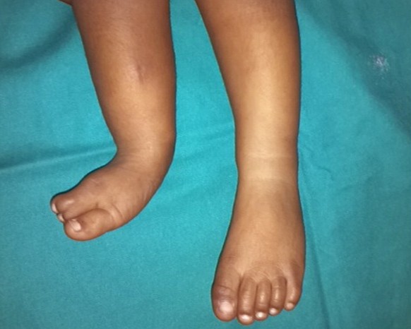
Picture of Fibular Hemimelia
Other factors have been incriminated in the appearance of fibular hemimelia. These factors are believed to lead to such congenital anomalies, affecting the development of the fetus between the 4th and 7th week of the pregnancy. Among these factors, there are: viral infection, trauma to the embryo, teratogenic environmental exposure and vascular dysgenesis. The latter is of particular importance, being represented by the failure of the embryo to form what should constitute a satisfactory blood supply.
Fibular Hemimelia Treatment
Fibular hemimelia requires surgical intervention in all cases. There are two main possibilities when it comes to such matters: a procedure to lengthen the affected limb (Ilizarov’s technique) or the amputation of the respective limb, followed by the application of a fitted prosthesis. In the majority of the patients, the amputation is performed around the age of 6 months. It is important to understand and remember is that the leg-lengthening procedures (also known as corrective osteotomies) are expensive. Moreover, they have to repeated a number of times, presenting an increased risk for residual deformity.
In choosing a particular treatment, the doctor will have to take into account the desire of the patient (or of the parents). There are people who are willing to go through repetitive, corrective surgeries rather than amputation, as they want to their lower limb to be as close to normal as it is possible. However, the possibility of amputation should be presented to the parents from the beginning.


 Cervix")
