
What is Angiokeratoma?
Also referred to as angiokeratomatas, this is a skin disease characterized by elevations from the skin accompanied by epidermal thickening.
- The prefix Angio – denotes blood or lymph vessels, a covering or an enclosure.
- Keratin is a protein found in the epidermis, the top most layer of the skin called the stratum corneum. It protects the deeper layers of the skin for example from excessive drying.
- The suffix – oma denotes a tumor either benign or malignant. Keratoma therefore refers to thick growths of the epidermis which may either be benign or malignant.
The dilated vessels may be caused by increased pressure within the veins. The increased pressure in the veins has been associated with:
- Varicocele
- Hernia
- Acute or chronic trauma
- Urinary system tumors
- Thrombophlebitis
- Vascular malformations
- Pregnancy
- Varicose veins in the vulva
- Post removal of the uterus
- Radiation therapy
However, not all patients with these conditions will develop angiokeratomas. Similarly not all patients with angiokeratomas will have these predisposing factors.
Angiokeratoma of Fordyce has been reported more in males, and are common in those who are older.
The angiokeratomas appear as raised areas on the skin (papules) and may appear as what is commonly known as a wart. They may appear in nay part of the body as described in the section on types of angiokeratomas.
Types of Angiokeratoma
There are several types of angiokeratomas, mainly classified according to the location. These include:
- Angiokeratoma of the scrotum and the vulva also known as Angiokeratoma of Fordyce. It can also be found at the shaft of the penis, labia majora inner thighs and the lower abdomen. The lesions may be small or large, red or blue/black. The surface may be scaly. They may also be may be solitary or multiple. Usually they do not cause any symptoms but may bleed easily when one scratches or after intercourse.
- Angiokeratoma circumsciptum (Verrucous vascular malformation) – usually a cluster of lesions on a small area of the body. Commonly found in the lower extremities and the lesions may darken in color over time. May be present at birth therefore some people call them “birthmarks”. This type presents with malformation of blood vessels underneath the epidermis
- Angiokeratoma corporis diffusum (Fabrys syndrome) – this is a rare form of the disease. It is genetic and more severe in males that females. The lesion are widespread, in the whole body with more distribution in the lower trunk and groin regions. It causes many complications including organ failure, blindness, painful hands, stroke and arthritis
- Sporadic angiokeratoma – involves solitary lesions on the skin common in persons over the age of 40 years. The lesions are small, bluish black papules and is common in lower extremities
- Mibellis angiokeratoma – described in 1889 by Mibelli , a type which affects the toes and fingers
Signs and Symptoms of Angiokeratoma
Generally angiokeratomas do not cause any symptoms that worry a patient. However the patient may note the following:
- Papules on the surface of the skin. The location may be scrotal skin, labia, glans penis, and shaft of the penis, abdomen, legs, fingers and upper thigh. Others include Angiokeratomas of the tongue, eyelid and vagina
- The lesion may be solitary or multiple
- Size of the lesions range form 1mm-5mm. younger patients tend to have smaller lesions and older patients have larger ones
- The color of the lesions may be red, blue or black. For younger patients, they tend to be red while older clients have darker lesions. Some small dilated veins may be visible beneath the skin.
- Bleeding from the lesions particularly those in the labia and scrotum may occur after intercourse or in pregnancy
- Itching and pain from the lesions is rare. Itching is reported more in the scrotum and the labia
- Overlying scales particularly in the older clients
Fabrys Syndrome however may have severe symptoms in males. These may include:
- Severe pains in the hands and feet
- Dark red skin rash in the umbilicus to the knees.
- Complications may result to
- Kidney failure
- Heart failure
- Neurological involvement
- Corneal opacity
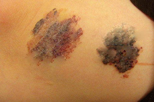
Angiokeratoma Pictures
Take a look at some of these pictures of Angiokeratoma:


Angiokeratoma Causes
The actual cause is unknown except for angiokeratoma corporis diffusum (Fabrys syndrome) which is genetic. It is an inherited disorder, passed by the X chromosome caused by deficiency of alpha-galactosidase enzyme. Lack of this enzyme causes deposition of glycosphingolipids in the inner lining of blood vessels (endothelium) and other areas for example the kidney, heart and nervous system.
Treatment for Angiokeratoma
Angiokeratomas are usually asymptomatic and do not cause any harm to the patient. In most cases there is no need of any form of treatment or medical care. However, since in the older patients they are blue/black in color and resemble skin cancer lesions, a biopsy may be performed to allay any anxiety and rule out malignant skin cancers.
- Where the angiokeratoma cause discomfort and bleeding, they may be removed surgically (excision).Clients may also request for their removal for cosmetic purposes.
- There are several surgical options available in the market today. Laser treatment has been proven to be effective.
- Other modalities of removal include cryotherapy or electrocautery. Cryotherapy involves the use liquid nitrogen or other gases to freeze the surface of the skin lesion.
- After surgical excision usually the healing is smooth with no complications.
- However Cryotherapy may result in a white mark or a scar noticeable in persons with a darker complexion, which can easily be concealed by applying appropriate makeup.
- If there is recurrence of the papules after surgical excision, follow-up is required to establish the actual type of lesion in order to rule out melanomas. Usually a biopsy is recommended.


 Cervix")
